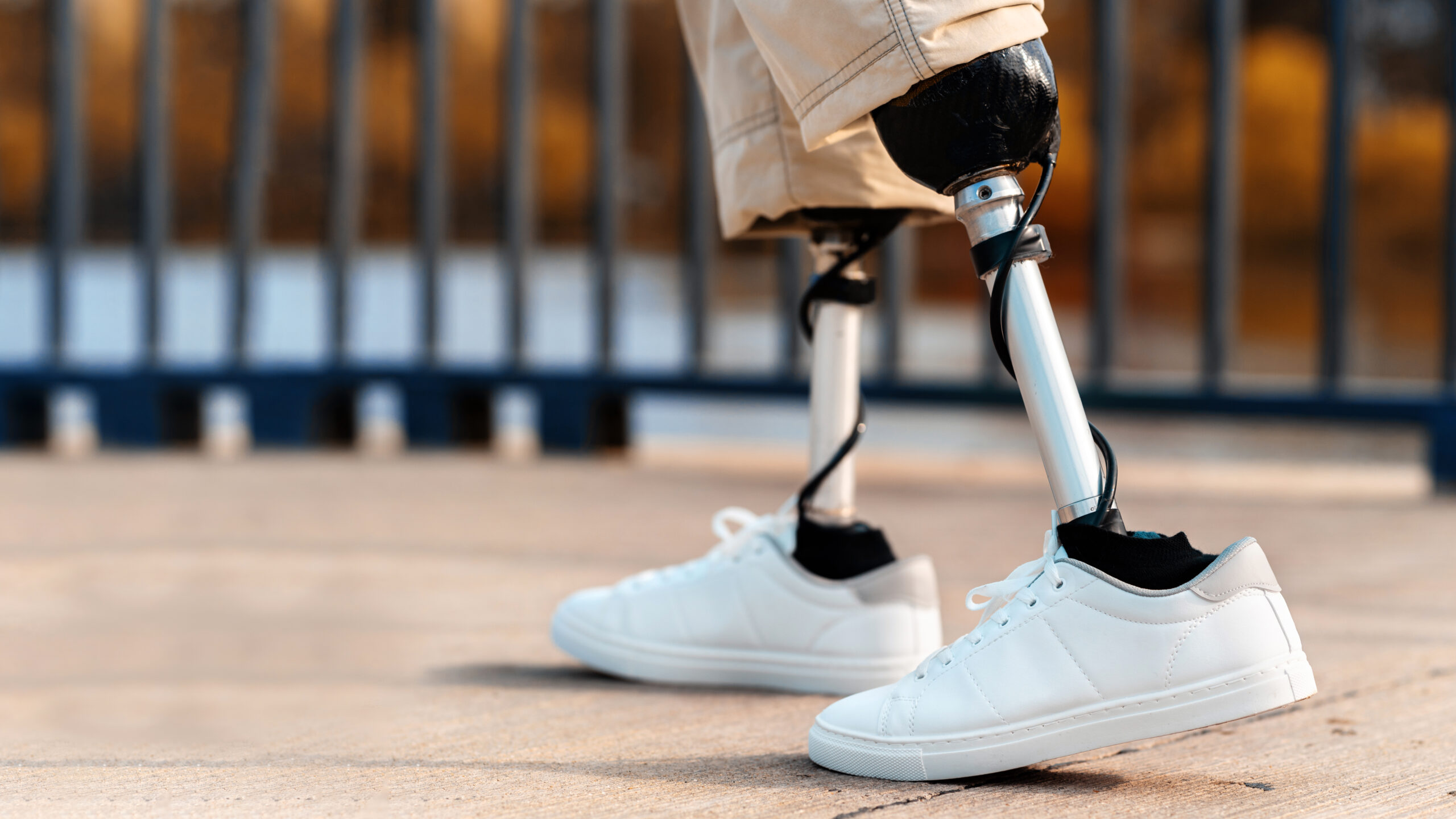
A new MIT study revealed a better method to control prosthetic devices – through magnetic beads. Currently, most prosthetic devices are controlled by electrodes. In an experimental study conducted at MIT, researchers replaced the electrodes with magnetic beads. Interestingly, this new system offered a more precise control of the connected prosthetic device.
Magnetomicrometry: New Approach To Control Prosthetic Devices
For those with a prosthetic device attached, the main challenge is controlling it to mimic natural movements. In most of the conventional configurations, prosthetic devices are controlled by electromyography – a technique to record the activity of the muscles.
Electrodes are either implanted in the muscles or inserted in the skin. These electrodes detect electric signals produced in the muscles when moved. These signals are then sent to the prosthesis, which begins to move, allowing the user to perform the desired actions. But this approach only offers limited control of the prosthesis.
According to Professor Hugh Herr of MIT, electromyography (EMG) does not provide an ideal solution. “When you use EMG-based control, you’re looking at an intermediate signal,” he explains. “You see what the brain is telling the muscle to do, but not what the muscle is actually doing.”
Instead, Professor Hugh Herr with his colleagues has developed a new technique named magnetomicrometry. They believe it could provide better controls than the traditional electromyography system. It involves two tiny magnetic beads inserted into the muscle tissues. These beads measure the length when muscles contract or expand and relay the information to a bionic prosthesis.
Promising Results So Far
The new system was tested on the calf muscles of turkeys. When the researchers moved the ankle joints of the bird, the magnetic sensors were able to accurately detect the movements associated with the joints. Most importantly, the detection time was 3 milliseconds – a significant improvement from EMG-based controls.
Two years ago professor Herr with his colleague, Cameron Taylor, a postdoctoral fellow at MIT, also another lead author of the study, developed an algorithm that reduced the time it takes for the system to detect the two magnetic beads. With this algorithm, they were able to tackle the long lead time that was the main obstacle for magnetomicrometry.
“With magnetomicrometry, we directly measure muscle length and speed,” says Herr. “Through mathematical modeling of the whole limb, we can calculate the positions and speeds of the prosthetic joints to be controlled, and then a simple robotic controller can control those joints.”
Professor Herr is optimistic that, clinical trials on humans could be conducted within few years. Furthermore, he adds that the technology could also be used in people with spinal cord injuries to restore mobility. Another advantage of magnetomicrometry is that it’s minimally invasive. Once implanted in the muscles, the magnetic beads will stay in place for a lifetime never needing another procedure or replacement.


